

AE-Adult-Echocardiography Free Certification Exam Easy to Download PDF Format 2026
Get 100% Success with Latest ARDMS RDCS AE-Adult-Echocardiography Exam Dumps
NEW QUESTION # 36
What is the direction of the mitral regurgitant jet in this video clip?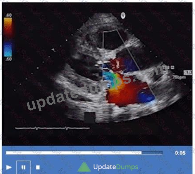
- A. Anterior
- B. Lateral
- C. Posterior
- D. Medial
Answer: C
Explanation:
The color Doppler jet in the apical four-chamber view is directed posteriorly toward the posterior left atrial wall, indicating a posteriorly directed mitral regurgitant jet.
Jet direction can provide insight into the mechanism of mitral regurgitation; for example, anterior leaflet prolapse often causes a posteriorly directed jet.
This analysis is outlined in the "Textbook of Clinical Echocardiography, 6e", Chapter on Mitral Regurgitation
- Jet Direction and Mechanism#20:390-395Textbook of Clinical Echocardiography#.
NEW QUESTION # 37
The respirometer should be turned on when assessing which possible disease process(es)?
- A. Congestive heart failure
- B. Ischemic cardiomyopathy
- C. Pericardial effusion and tamponade
- D. Mitral regurgitation and stenosis
Answer: C
Explanation:
A respirometer monitors the respiratory cycle and is essential when evaluating diseases in which respiratory variation affects echocardiographic measurements, such as pericardial effusion and cardiac tamponade. In tamponade, respiratory changes in mitral and tricuspid inflows, as well as variations in inferior vena cava size, are key diagnostic features.
Congestive heart failure, ischemic cardiomyopathy, and mitral valve diseases do not require synchronization with respiration for diagnosis or quantification and are not reliant on respirometer use.
This recommendation is outlined in ASE pericardial disease guidelines and echocardiography procedural protocols#16:Textbook of Clinical Echocardiography, 6ep.280-285##12:ASE Pericardial Disease Guidelinesp.300-305#.
NEW QUESTION # 38
Which acute disease state is indicated with McConnell's sign?
- A. Pulmonary embolism
- B. Aortic dissection
- C. Libman-Sacks endocarditis
- D. Myocardial infarction
Answer: A
Explanation:
McConnell's sign is an echocardiographic finding characterized by regional right ventricular (RV) dysfunction with akinesia of the mid-free wall but preserved contractility of the apex. This pattern is highly specific for acute pulmonary embolism (PE).
In acute PE, sudden obstruction of the pulmonary artery leads to acute right ventricular pressure overload, causing regional wall motion abnormalities. The sparing of the apex differentiates it from other causes of RV dysfunction such as myocardial infarction.
This sign is considered a useful bedside clue in the echocardiographic diagnosis of PE, especially when combined with clinical findings and Doppler evidence of elevated pulmonary pressures.
The sign is described in the "Textbook of Clinical Echocardiography, 6e", Chapter on Acute Right Heart Dysfunction, with reference to McConnell's original description and its clinical significance in acute pulmonary embolism diagnosis#20:340-345Textbook of Clinical Echocardiography#.
NEW QUESTION # 39
Which structure is the arrow pointing to in this video?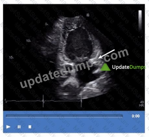
- A. Left lower pulmonary vein
- B. Left atrial appendage
- C. Descending aorta
- D. Coronary sinus
Answer: D
Explanation:
The arrow points to the coronary sinus, which is a venous structure located posteriorly in the atrioventricular groove, emptying into the right atrium. It appears as a circular anechoic structure near the left atrium in echocardiographic images.
Left lower pulmonary vein enters the left atrium more superiorly. Descending aorta is posterior to the heart but not in this location. Left atrial appendage is an anterior finger-like projection of the left atrium, separate from the coronary sinus.
This anatomy is described in the "Textbook of Clinical Echocardiography, 6e", Chapter on Cardiac Venous Anatomy#20:140-145Textbook of Clinical Echocardiography#.
NEW QUESTION # 40
Which of the following does the pulmonary capillary wedge pressure estimate?
- A. Right ventricular pressure
- B. Left ventricular pressure
- C. Right atrial pressure
- D. Left atrial pressure
Answer: D
Explanation:
Comprehensive and Detailed Explanation From Exact Extract:
Pulmonary capillary wedge pressure (PCWP) is obtained by advancing a balloon-tipped catheter into a small branch of the pulmonary artery and inflating the balloon to "wedge" the catheter, thereby occluding forward blood flow and measuring the pressure distal to the occlusion. The measured pressure reflects the pressure in the pulmonary venous system, which closely approximates left atrial pressure (LAP) under normal conditions.
Since the left atrium receives pulmonary venous return before the blood enters the left ventricle, PCWP is a surrogate for LAP, which in turn reflects left ventricular end-diastolic pressure (LVEDP) in the absence of mitral valve disease or pulmonary venous obstruction. PCWP is widely used in clinical and echocardiographic contexts to estimate left heart filling pressures.
It does not estimate right atrial, right ventricular, or left ventricular pressures directly. Right atrial pressure is measured via central venous pressure, right ventricular pressure by catheterization, and left ventricular pressure by direct catheterization.
This concept is extensively discussed in the "Textbook of Clinical Echocardiography, 6e", Chapter on Hemodynamics and Doppler Assessment, with specific emphasis on the use of PCWP to estimate left atrial pressure#20:200-210Textbook of Clinical Echocardiography#.
NEW QUESTION # 41
Which is most likely the culprit coronary artery in a patient who presents with anteroseptal hypokinesis?
- A. Posterior descending artery
- B. Right coronary artery
- C. Circumflex artery
- D. Left coronary artery
Answer: D
Explanation:
Comprehensive and Detailed Explanation From Exact Extract:
Anteroseptal hypokinesis is most often due to ischemia or infarction in the left anterior descending (LAD) artery territory, a major branch of the left coronary artery. The LAD supplies the anterior wall and the interventricular septum.
The right coronary artery generally supplies the inferior wall and right ventricle. The circumflex artery supplies the lateral wall. The posterior descending artery supplies the inferior wall.
This coronary artery distribution and wall motion correlation is fundamental in stress echocardiography and ischemic heart disease assessment as detailed in ASE guidelines and clinical echocardiography references#12:
ASE Stress Echocardiography Guidelinesp.300-310##16:Textbook of Clinical Echocardiography, 6ep.380-
385#.
NEW QUESTION # 42
Which mitral valve filling pattern is characterized by a long deceleration time and an E/A ratio of 0.6?
- A. Impaired relaxation
- B. Normal
- C. Restrictive
- D. Pseudonormal
Answer: A
Explanation:
The mitral valve filling pattern characterized by a long deceleration time and a reduced E/A ratio (less than 1, such as 0.6) is consistent with impaired relaxation. This pattern is typically seen in early diastolic dysfunction, where there is slowed ventricular relaxation, resulting in reduced early diastolic filling (E wave) and a compensatory increase in atrial contraction contribution (A wave).
Impaired relaxation pattern shows:
E/A ratio < 1 (e.g., 0.6)
Prolonged deceleration time (>200 ms)
Prolonged isovolumic relaxation time (IVRT)
This pattern differs from restrictive filling, which has a high E/A ratio (>2), shortened deceleration time (<150 ms), and elevated left atrial pressures. Pseudonormal filling has a normal or near-normal E/A ratio but elevated filling pressures that mask underlying dysfunction and requires further evaluation with tissue Doppler or pulmonary venous flow for diagnosis. Normal filling has a typical E/A ratio around 1 to 1.5 with normal deceleration times.
The textbook details that impaired relaxation is the earliest sign of diastolic dysfunction and describes the prolongation of the deceleration time and reduced E/A ratio as hallmark findings of this stage.
NEW QUESTION # 43
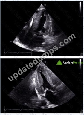
Which diagnosis is most consistent with the findings in these images?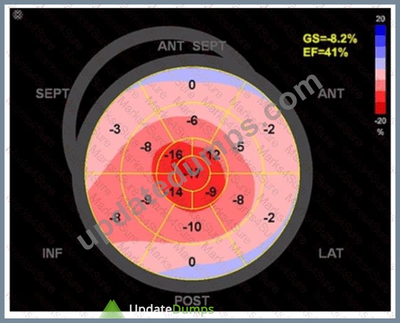
- A. Hypertrophic obstructive cardiomyopathy
- B. Restrictive cardiomyopathy from amyloidosis
- C. Apical hypertrophic cardiomyopathy
- D. Takotsubo cardiomyopathy
Answer: D
Explanation:
The first image shows a bullseye plot of global longitudinal strain (GLS) with marked reduction in strain values (less negative numbers) most prominently in the apical segments (central red zone), with an overall GLS of -8.2% (normal is about -20%) and a reduced ejection fraction of 41%. This pattern is characteristic of Takotsubo cardiomyopathy, which typically demonstrates regional wall motion abnormalities that predominantly involve the apex and mid segments of the left ventricle with basal sparing.
The 2D echocardiographic images show apical ballooning, a hallmark of Takotsubo cardiomyopathy, where the apex is akinetic or dyskinetic and the basal segments contract normally or hypercontract. Doppler images show findings consistent with impaired ventricular function.
In contrast:
Apical hypertrophic cardiomyopathy (HCM) would show increased wall thickness localized to the apex but not apical ballooning or reduced strain in that typical pattern.
Hypertrophic obstructive cardiomyopathy (HOCM) involves basal septal hypertrophy with outflow obstruction, not apical akinesis or ballooning.
Restrictive cardiomyopathy from amyloidosis involves diffuse infiltration and generally a different strain pattern with more uniform reduction and "apical sparing" rather than apical involvement.
This interpretation aligns with the diagnostic criteria and echocardiographic features described in the adult echocardiography literature, including the "Textbook of Clinical Echocardiography" (Chapter on Cardiomyopathies) and ASE guidelines, which highlight apical ballooning and regional strain abnormalities as diagnostic features of Takotsubo cardiomyopathy#16:Cardiomyopathy ChapterTextbook of Clinical Echocardiography, 6e##12:ASE Guidelines on Strain Imagingp.130-135#.
NEW QUESTION # 44
When utilizing contrast agents, what should the sonographer keep in mind?
- A. Contrast agents produce only mild reactions.
- B. Patients with no history of allergies will not have reactions.
- C. The contrast-enhanced exam poses no risk to the patient.
- D. Anaphylactoid reactions may occur.
Answer: D
Explanation:
Contrast agents used in echocardiography can rarely cause anaphylactoid reactions, which are non-IgE- mediated hypersensitivity reactions that can mimic anaphylaxis. Therefore, sonographers must be prepared to manage such reactions.
Contrary to option B, reactions can be severe though rare. Even patients without prior allergies can react. It is incorrect to say the exam poses no risk; proper precautions and monitoring are essential.
These precautions are emphasized in ASE contrast echocardiography guidelines and safety protocols#12:ASE Contrast Echocardiography Guidelinesp.190-195##16:Textbook of Clinical Echocardiography, 6ep.575-
580#.
NEW QUESTION # 45
Which type of defect can be seen in this video clip?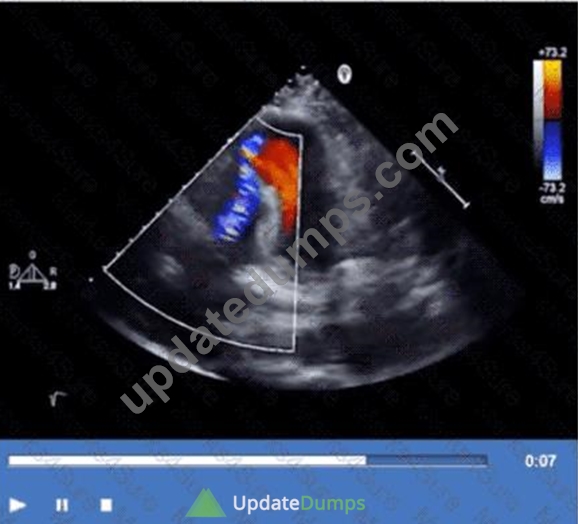
- A. Pseudoaneurysm of the apex
- B. Tricuspid regurgitation
- C. Ischemic ventricular septal defect
- D. Coronary artery aneurysm
Answer: C
Explanation:
The echocardiographic video shows a defect in the ventricular septum with left-to-right shunting consistent with an ischemic ventricular septal defect (VSD), a mechanical complication of myocardial infarction. The defect allows blood flow between the left and right ventricles.
Coronary artery aneurysm appears as dilated coronary vessels, not a septal defect. Tricuspid regurgitation involves the right atrioventricular valve and is identified differently. Pseudoaneurysm of the apex is a contained myocardial rupture with narrow neck and does not involve septal communication.
This complication and its echocardiographic features are described in the "Textbook of Clinical Echocardiography, 6e", Chapter on Post-Infarction Mechanical Complications#20:430-435Textbook of Clinical Echocardiography#.
NEW QUESTION # 46
Which of the following can be calculated from the peak tricuspid regurgitant velocity?
- A. Pulmonary artery diastolic pressure
- B. Right ventricular systolic pressure
- C. Mean pulmonary artery pressure
- D. Right atrial pressure
Answer: B
Explanation:
Peak tricuspid regurgitant velocity (TRV) allows estimation of right ventricular systolic pressure (RVSP) using the simplified Bernoulli equation: RVSP = 4 × (TRV)^2 + estimated right atrial pressure.
This measurement is important for assessing pulmonary hypertension indirectly.
Right atrial pressure is estimated separately, pulmonary artery diastolic pressure and mean pressure require additional measurements.
This application is discussed in the "Textbook of Clinical Echocardiography, 6e", Chapter on Right Heart Pressure Estimation#20:335-340Textbook of Clinical Echocardiography#.
NEW QUESTION # 47
Which view best demonstrates a wall thickening abnormality of the apical lateral segment?
- A. Four-chamber
- B. Mid-parastemal short axis
- C. Parasternal long axis
- D. Two-chamber
Answer: D
Explanation:
The two-chamber apical view allows visualization of the left ventricle's anterior and inferior walls, including the apical lateral segment. It is ideal for assessing wall thickness and segmental wall motion abnormalities in this region.
The four-chamber view visualizes septal and lateral walls but does not optimally display the apical lateral segment. Parasternal long axis primarily visualizes the anterior septum and posterior wall but is limited for lateral apex. The mid-parasternal short axis focuses on mid-ventricular segments and does not visualize the apex.
This anatomical and echocardiographic detail is described in the "Textbook of Clinical Echocardiography,
6e", Chapter on Left Ventricular Segmental Analysis#20:120-125Textbook of Clinical Echocardiography#.
NEW QUESTION # 48
Which parameter is necessary to calculate a 2D left atrial volume index?
- A. Cardiac output
- B. Age
- C. Height
- D. Blood pressure
Answer: C
Explanation:
Comprehensive and Detailed Explanation From Exact Extract:
The left atrial volume index (LAVI) is the left atrial volume normalized to the patient's body surface area (BSA), which accounts for patient size. To calculate BSA, height and weight are required, most commonly using formulas such as the Mosteller formula.
Therefore, height is a necessary parameter to calculate the left atrial volume index. Age, blood pressure, and cardiac output are not used in the calculation of LAVI but may be clinically relevant for interpretation.
This approach standardizes LA size across patients of different body habitus, making LAVI a more accurate and reproducible measure of LA remodeling and a predictor of cardiovascular outcomes.
The echocardiography guidelines and textbooks emphasize the importance of indexing LA volume to BSA and highlight height as a required measurement for this purpose .
NEW QUESTION # 49
In patients with interrupted aortic arch, which structure allows Wood to flow into the descending aorta?
- A. Left carotid artery
- B. Persistent left superior vena cava
- C. Patent ductus arteriosus
- D. Foramen ovale
Answer: C
Explanation:
In interrupted aortic arch, the normal continuity between the ascending and descending aorta is disrupted. The patent ductus arteriosus (PDA) provides a vital conduit for blood to flow from the pulmonary artery to the descending aorta, maintaining systemic circulation distal to the interruption.
Persistent left superior vena cava and left carotid artery do not provide this flow. The foramen ovale is an atrial-level shunt and does not compensate for interrupted aortic arch.
This clinical anatomy is described in the "Textbook of Clinical Echocardiography, 6e", Chapter on Congenital Aortic Arch Anomalies#20:135-140Textbook of Clinical Echocardiography#.
NEW QUESTION # 50
In cardiac tamponade, how do transvalvular pressure gradients change during expiration?
- A. Transmitral decreases and transtricuspid increases
- B. Transmitral increases and transtricuspid increases
- C. Transmitral decreases and transtricuspid decreases
- D. Transmitral increases and transtricuspid decreases
Answer: A
Explanation:
In cardiac tamponade, there is a characteristic reciprocal respiratory variation in transvalvular flow velocities due to ventricular interdependence and impaired cardiac filling. During expiration, the intrathoracic pressure increases, which leads to decreased right ventricular filling and thus decreased transtricuspid flow velocity.
Simultaneously, left ventricular filling increases, causing an increase in transmitral flow velocity.
Therefore, during expiration, the transmitral gradient increases while the transtricuspid gradient decreases.
This phenomenon reverses during inspiration, where transtricuspid flow increases and transmitral flow decreases. These respiratory variations are diagnostic hallmarks of tamponade physiology and help distinguish it from other conditions.
This principle is illustrated in Doppler echocardiographic studies of ventricular inflow and is described with diagrams in the "Textbook of Clinical Echocardiography, 6e" (Chapter 10: Pericardial Disease), highlighting the changes in transmitral and transtricuspid velocities during the respiratory cycle in tamponade .
NEW QUESTION # 51
Which diagnosis is most likely confirmed by echocardiography in a 65-year-old female presenting with new onset chest pain associated with ST segment elevation on the electrocardiogram and angiographically normal coronary artenes?
- A. Restrictive cardiomyopathy
- B. Alcohol-associated cardiomyopathy
- C. Apical hypertrophic cardiomyopathy
- D. Takotsubo cardiomyopathy
Answer: D
Explanation:
Takotsubo cardiomyopathy, also known as stress-induced cardiomyopathy or "broken heart syndrome," predominantly affects postmenopausal women (usually older than 50 years) and often presents with acute chest pain and ST-segment elevation on the ECG mimicking acute myocardial infarction. However, coronary angiography reveals normal or non-obstructive coronary arteries.
Echocardiographically, Takotsubo cardiomyopathy is characterized by transient left ventricular systolic dysfunction with a typical pattern of apical ballooning and basal hyperkinesis. The wall motion abnormality extends beyond a single coronary artery territory, differentiating it from ischemic cardiomyopathy.
The diagnosis is supported by the clinical presentation, typical echocardiographic findings, and exclusion of obstructive coronary artery disease. The condition is usually reversible over days to weeks.
This is extensively described in the "Textbook of Clinical Echocardiography, 6e" (Chapter 8: Coronary Artery Disease and Takotsubo Syndrome), which highlights the typical patient demographics, presentation, echocardiographic features, and prognosis .
NEW QUESTION # 52
......
Get Ready to Pass the AE-Adult-Echocardiography exam Right Now Using Our ARDMS RDCS Exam Package: https://www.updatedumps.com/ARDMS/AE-Adult-Echocardiography-updated-exam-dumps.html
The Best AE-Adult-Echocardiography Exam Study Material and Preparation Test Question Dumps: https://drive.google.com/open?id=1DrPyp_HkorGNN07LIju53GERkrmfnYkR